Breast cancer is the most common female cancer in the US.
Important
risk factors for breast cancer are age, gender, reproductive history, hormonal
factors, and family history. Although a family history of breast and/or ovarian
cancer is common in patients diagnosed with breast cancer, less than ten
percent of all breast cancers are associated with genetic mutations.
Assay
of hormone receptors (estrogen [ER] and progesterone [PR] receptors) is an
important component of the pathologic evaluation of breast cancer, for both
prognostic and predictive purposes, as patients with hormone receptor-positive
tumors benefit from the addition of endocrine treatments.
What are the Molecular SubTypes of Breast Cancer:
Molecular profiling,
based upon variations in gene expression, has identified several distinct
breast cancer subtypes, named the breast cancer intrinsic subtypes.
Luminal A and B are the most common subtypes and
make up the majority of estrogen receptor (ER)-positive breast cancers. Luminal
A tumors carry the best prognosis of all of the subtypes. Luminal B tumors have
a lower expression of ER-related genes, variable expression of the human
epidermal growth factor receptor-2 (HER2) cluster, and higher expression of the
proliferation cluster. Consequently, luminal B tumors have a poorer prognosis.
HER2-enriched (HER2+/ER-) subtype cancers are
typically negative for ER and progesterone receptor (PR), and positive for
HER2. The poor prognosis associated with these tumors has been undoubtedly
altered by HER2-directed therapies
Basal-like tumors are typically ER-negative,
PR-negative, and HER2-negative on clinical assays, which has prompted the
nickname "triple-negative" to describe them. They have a strong
association with cancers arising in breast cancer gene 1, early onset (BRCA1)
mutation carriers and are overrepresented in breast cancer developing during the
premenopausal years and in African-American women. They have the worst
prognosis of the subtypes
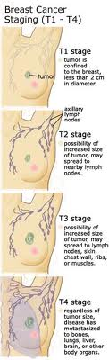
Staging of Breast
Cancer:
Tumor node metastases (TNM) staging system for carcinoma of the
breast
Primary tumor (T)
TX
Primary
tumor cannot be assessed
T0
No
evidence of primary tumor
Tis
Carcinoma
in situ
Tis
(DCIS)
Ductal
carcinoma in situ
Tis
(LCIS)
Lobular
carcinoma in situ
Tis
(Paget's)
Paget's
disease (Paget disease) of the nipple NOT associated with invasive carcinoma
and/or carcinoma in situ (DCIS and/or LCIS) in the underlying breast
parenchyma. Carcinomas in the breast parenchyma associated with Paget's
disease are categorized based on the size and characteristics of the
parenchymal disease, although the presence of Paget's disease should still be
noted.
T1
Tumor
≤20 mm in greatest dimension
T1mi
Tumor
≤1 mm in greatest dimension
T1a
Tumor
>1 mm but ≤5 mm in greatest dimension
T1b
Tumor
>5 mm but ≤10 mm in greatest dimension
T1c
Tumor
>10 mm but ≤20 mm in greatest dimension
T2
Tumor
>20 mm but ≤50 mm in greatest dimension
T3
Tumor
>50 mm in greatest dimension
T4◊
Tumor
of any size with direct extension to the chest wall and/or to the skin
(ulceration or skin nodules)
T4a
Extension
to the chest wall, not including only pectoralis muscle adherence/invasion
T4b
Ulceration
and/or ipsilateral satellite nodules and/or edema (including peau d'orange)
of the skin, which do not meet the criteria for inflammatory carcinoma
T4c
Both
T4a and T4b
T4d
Inflammatory
carcinoma§
The
use of neoadjuvant therapy does not change the clinical (pretreatment) stage.
Clinical (pretreatment) T will be defined by clinical and radiographic
findings, while y pathologic (posttreatment) T will be determined by
pathologic size and extension. The ypT will be measured as the largest single
focus of invasive tumor, with the modifier "m" indicating multiple
foci. The measurement of the largest tumor focus should not include areas of
fibrosis within the tumor bed.
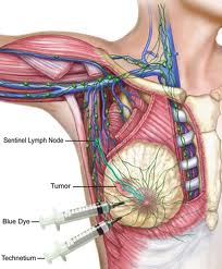
Regional lymph
nodes (N)
Clinical
NX
Regional
lymph nodes cannot be assessed (eg, previously removed)
N0
No
regional lymph node metastases
N1
Metastases
to movable ipsilateral level I, II axillary lymph node(s)
N2
Metastases
in ipsilateral level I, II axillary lymph nodes that are clinically fixed or
matted; or in clinically detected‡ ipsilateral internal mammary nodes in theabsenceof clinically
evident axillary lymph node metastases
N2a
Metastases
in ipsilateral level I, II axillary lymph nodes fixed to one another (matted)
or to other structures
N2b
Metastases
only in clinically detected‡ ipsilateral internal mammary nodes and in theabsenceof clinically
evident level I, II axillary lymph node metastases
N3
Metastases
in ipsilateral infraclavicular (level III axillary) lymph node(s) with or
without level I, II axillary lymph node involvement; or in clinically
detected‡ ipsilateral internal mammary lymph node(s) with clinically evident
level I, II axillary lymph node metastases; or metastases in ipsilateral
supraclavicular lymph node(s) with or without axillary or internal mammary
lymph node involvement
N3a
Metastases
in ipsilateral infraclavicular lymph node(s)
N3b
Metastases
in ipsilateral internal mammary lymph node(s) and axillary lymph node(s)
N3c
Metastases
in ipsilateral supraclavicular lymph node(s)
Pathologic
(pN)†**
pNX
Regional
lymph nodes cannot be assessed (eg, previously removed, or not removed for
pathologic study)
pN0
No
regional lymph node metastasis identified histologically
pN0(i-)
No
regional lymph node metastases histologically, negative immunohistochemistry
(IHC)
pN0(i+)
Malignant
cells in regional lymph node(s) no greater than 0.2 mm (detected by H&E
or IHC including isolated tumor cell clusters (ITC))
pN0(mol-)
No
regional lymph node metastases histologically, negative molecular findings
(RT-PCR)••
pN0(mol+)
Positive
molecular findings (RT-PCR)••, but no regional lymph node metastases detected
by histology or IHC
pN1
Micrometastases;
or metastases in 1-3 axillary lymph nodes; and/or in internal mammary nodes
with metastases detected by sentinel lymph node biopsy but not clinically
detected ΔΔ
pN1mi
Micrometastases
(greater than 0.2 mm and/or more than 200 cells, but none greater than 2.0
mm)
pN1a
Metastases
in 1-3 axillary lymph nodes, at least one metastasis greater than 2.0 mm
pN1b
Metastases
in internal mammary nodes with micrometastases or macrometastases detected by
sentinel lymph node biopsy but not clinically detected ΔΔ
pN1c
Metastases
in 1-3 axillary lymph nodes and in internal mammary lymph nodes with
micrometastases or macrometastases detected by sentinel lymph node biopsy but
not clinically detected
pN2
Metastases
in 4-9 axillary lymph nodes; or in clinically detected◊◊ internal mammary
lymph nodes in theabsenceof axillary lymph
node metastases
pN2a
Metastases
in 4-9 axillary lymph nodes (at least one tumor deposit greater than 2.0 mm)
pN2b
Metastases
in clinically detected◊◊ internal mammary lymph nodes in theabsenceof axillary lymph
node metastases
pN3
Metastases
in ten or more axillary lymph nodes; or in infraclavicular (level III
axillary) lymph nodes; or in clinically detected◊◊ ipsilateral internal
mammary lymph nodes in thepresenceof one or more
positive level I, II axillary lymph nodes; or in more than three axillary
lymph nodes and in internal mammary lymph nodes with micrometastases or
macrometastases detected by sentinel lymph node biopsy but not clinically
detected ΔΔ; or in ipsilateral supraclavicular lymph nodes
pN3a
Metastases
in ten or more axillary lymph nodes (at least one tumor deposit greater than
2.0 mm); or metastases to the infraclavicular (level III axillary lymph)
nodes
pN3b
Metastases
in clinically detected◊◊ ipsilateral internal mammary lymph nodes in thepresenceof one or more
positive axillary lymph nodes; or in more than three axillary lymph nodes and
in internal mammary lymph nodes with micrometastases or macrometastases
detected by sentinel lymph node biopsy but not clinically detected ΔΔ
pN3c
Metastases
in ipsilateral supraclavicular lymph nodes
Posttreatment
ypN
-
Post-treatment yp "N" should be evaluated as for clinical
(pretreatment) "N" methods above. The modifier "sn" is
used only if a sentinel node evaluation was performed after treatment. If no
subscript is attached, it is assumed that the axillary nodal evaluation was
by axillary node dissection (AND).
-
The X classification will be used (ypNX) if no yp posttreatment SN or AND was
performed
-
N categories are the same as those for pN
Distant metastasis
(M)
M0
No
clinical or radiographic evidence of distant metastases
cM0(i+)
No
clinical or radiographic evidence of distant metastases, but deposits of
molecularly or microscopically detected tumor cells in circulating blood,
bone marrow, or other nonregional nodal tissue that are no larger than 0.2 mm
in a patient without symptoms or signs of metastases
M1
Distant
detectable metastases as determined by classic clinical and radiographic
means and/or histologically proven larger than 0.2 mm
Posttreatment yp M classification.The M category for
patients treated with neoadjuvant therapy is the category assigned in the
clinical stage, prior to initiation of neoadjuvant therapy. Identification of
distant metastases after the start of therapy in cases where pretherapy
evaluation showed no metastases is considered progression of disease. If a
patient was designated to have detectable distant metastases (M1) before
chemotherapy, the patient will be designated as M1 throughout.
Anatomic
stage/prognostic groups§§
0
Tis
N0
M0
IA
T1
¥¥
N0
M0
IB
T0
N1mi
M0
T1
¥¥
N1mi
M0
IIA
T0
N1
‡‡
M0
T1
¥¥
N1
‡‡
M0
T2
N0
M0
IIB
T2
N1
M0
T3
N0
M0
IIIA
T0
N2
M0
T1
¥¥
N2
M0
T2
N2
M0
T3
N1
M0
T3
N2
M0
IIIB
T4
N0
M0
T4
N1
M0
T4
N2
M0
IIIC
Any
T
N3
M0
IV
Any
T
Any
N
M1
.
Treatment of hormone receptor positive breast cancer:
The
treatment of early stage breast cancer includes the treatment of locoregional
disease with surgery, radiation therapy, or both, and the treatment of systemic
disease with one or a combination of chemotherapy, endocrine therapy, or
biologic therapy.
Neoadjuvant (before
Surgery) systemic therapy has become a frequently used option in the treatment
of breast cancer.
For patients with locally advanced, inoperable
breast cancer and inflammatory breast cancer, neoadjuvant systemic therapy is
standard treatment. The goal of neoadjuvant systemic therapy is to induce tumor
response, to facilitate local control through surgical resection and radiation
therapy, and to improve disease-free and overall survival.
For patients with early stage, operable breast
cancer, neoadjuvant systemic therapy may be used rather than primary surgery in
order to increase the chance of successful breast conserving surgery. For these
patients, neoadjuvant systemic therapy results in long-term distant
disease-free survival and overall survival comparable to that achieved with
adjuvant systemic therapy.
Here,
Dr. Tony Talebi discusses the general concepts of estrogen receptor positive
breast cancer with world renowned breast cancer expert Dr. Marc Lippman, professor
and chairman of the department of medicine at the University of Miami. Dr. Marc
Lippman pioneered the use of tamoxifen in estrogen receptor positive breast
cancer with his early research while at the National Cancer Institute which
revolutionized the treatment of breast cancer. The discussion
includes breast
cancer stage treatment, advanced breast cancer treatment, symptom of breast
cancer, new treatments for breast cancer, stage 1 breast cancer treatment,
stage 4 cancer treatment, early symptoms of breast cancer, early symptoms of
breast cancer, staging of breast cancer, symptoms for breast cancer, stage iv
breast cancer treatment, breast cancer cells, men breast cancer, breast cancer
in women, risk factors of breast cancer, radiation for breast cancer,
chemotherapy for breast cancer, breast cancer radiation, advanced cancer
treatment, what causes breast cancer, articles on breast cancer, facts on
breast cancer, stage 3 breast cancer, risk factors for breast cancer, symptoms
of cancer, breast cancer survival rates, what cause breast cancer, breast
cancer risk factors, stage 3 breast cancer prognosis, survival rate for breast
cancer, early stage breast cancer, images of breast cancer, inflammatory breast
cancer symptoms, how is breast cancer diagnosed, breast cancer holistic
treatment, triple negative breast cancer prognosis, triple negative breast
cancer survival, effects of breast cancer, holistic cancer treatment, different
types of breast cancer, first signs of breast cancer, stage 4 breast cancer prognosis,
definition of breast cancer, statistics of breast cancer.
Dr. Marc Lippman credentials:
Certifications
American Board of Internal Med-Medical Oncology
American Board of Internal Medicine
American Bd of Int Med-Endocrinology Diabetes & Metabolism
Specialties
Hematology/Oncology - Internal Medicine
Internal Medicine
Roles
Interim Deputy Director, Sylvester Comprehensive Cancer Center
Professor and Chairman
Biography
Marc E. Lippman, M.D. was named the Kathleen and Stanley Glaser Professor of Medicine at the University of Miami Leonard M. Miller School of Medicine, and was named Chairman of the Department of Medicine in May 2007. Previously Dr. Lippman was the John G. Searle Professor and Chair of Internal Medicine at the University of Michigan, Ann Arbor, Michigan. From 1988 through 1999 Dr. Lippman was Professor of Medicine and Pharmacology, and Chair, Department of Oncology at Georgetown University in Washington, D.C., and served as Director of the Lombardi Cancer Center at Georgetown University Medical Center. From 1978 through 1990 he was Clinical Professor of Medicine and Pharmacology, Uniformed Services, University of the Health Sciences. Dr. Lippman served as Head of the Medical Breast Cancer Section, Medicine Branch, at the National Institute of Health. He was a Senior Investigator at the National Cancer Institute of the National Institute of Health. Dr. Lippman completed a Fellowship in Endocrinology at Yale Medical School in New Haven, CT from 1973-1974. In addition, he was Clinical Associate at the National Cancer Institute from 1970-1971 and Clinical Associate at the Laboratory of Biochemistry of the National Cancer Institute of the National Institute of Health. From 1970 to 1988 he served as an Officer and Medical Director of the United States Public Health Service. Dr. Lippman completed his residency on the Osler Medical Service, John Hopkins Hospital, in Baltimore, Maryland from 1968-1970.
A native of New York, Dr. Lippman received his Bachelor's Degree from Cornell University, Magna Cum Laude, and medical school degree at Yale Medical School in New Haven, CT where he was elected to AOA.
Dr. Lippman is widely known for his research in breast cancer. Throughout his illustrious career he has received numerous awards including Mallinckrodt Award of the Clinical Radioassay Society in 1978; the Commendation Medal USPHS in 1982; Meritorius Service Medal, USPHS in 1987; Clinical Investigator Award, American Federation for Clinical Research in 1985; D.R. Edwards Lecture and Medal, Tenovus Institute, Wales 1985; Plenary Lecturer, British Association of Cancer Research in 1987; Gosse Lecture, Dalhuosie University, Halifax Nova Scotia in 1987; the American Cancer Society.