How is the MUGA scan performed?
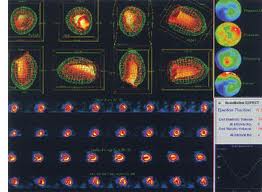
A MUGA scan is performed by attaching a radioactive substance, Technetium 99, to red blood cells, then injecting the red blood cells into the patient’s bloodstream. The patient is then placed under a special camera (a gamma camera), which is able to detect the low-level radiation being given off by the Technetium-labelled red cells. Since the red blood cells (including those that are radio-labelled) fill the cardiac chambers, the image produced by the gamma camera is essentially an outline of those chambers. With some fancy computer manipulation, the the final product is a movie of the heart beating.What can be learned from the MUGA scan?
For the purposes of hematology/oncology, the MUGA scan gives an accurate and reproducible means of measuring and monitoring the ejection fraction of the cardiac ventricles. Since some of the chemotherpies we utilize can be toxic to the heart, we monitor the heart function with checking MUGA scans before, during and after certain chemotherpies which are potentially toxic to the heart muscles.The left ventricular ejection fraction (LVEF) is an excellent, and the most commonly used, measure of overall cardiac function. The ejection fraction is simply the proportion of blood that is expelled from the ventricle with each heart beat. So, for instance, if the left ventricle ejects 60% of its blood volume with each beat, the LVEF is 0.6. (A normal LVEF is 0.5 or greater.)